
Every day in emergency rooms and urgent care centers across Florida, patients arrive with lower back pain, leg weakness, or new bladder symptoms — and the way their doctor responds to those complaints can determine whether they walk out of the hospital or spend the rest of their life managing permanent disability.
Cauda Equina Syndrome (CES) is one of the most time-critical conditions in medicine. It is also one of the most commonly misdiagnosed. The reasons for that are not mysterious — lower back pain is one of the most common complaints in any clinical setting, and the early symptoms of CES overlap with far more common and far less urgent conditions like herniated discs, sciatica and lumbar strain. But while those conditions allow time for conservative management and watchful waiting, CES does not. It demands immediate, decisive action.
Understanding how CES is supposed to be diagnosed — what a proper clinical workup looks like, what tests must be ordered, and what findings must not be dismissed — is essential for patients who suspect they received substandard care. It is also the foundation of every CES medical malpractice case we handle.
If you are already dealing with the aftermath of a delayed diagnosis and need legal guidance, learn more on our main Florida Cauda Equina Syndrome Lawyer page.
What Causes Cauda Equina Syndrome
The cauda equina is the bundle of nerve roots at the base of the spinal cord — named for their resemblance to a horse’s tail — that branch off below the L1-L2 vertebrae and travel downward through the lumbar and sacral spine. These nerves carry the motor and sensory signals that control the legs, bladder, bowel and sexual function.
CES occurs when those nerve roots are compressed, disrupting the signals they carry. The most common causes include:
Lumbar Disc Herniation: A large herniated disc at the L4-L5 or L5-S1 level is responsible for the majority of CES cases — accounting for approximately 45 percent of all presentations. A disc that herniates centrally and severely can compress multiple nerve roots simultaneously, triggering the full CES picture rapidly.
Spinal Stenosis: Narrowing of the spinal canal, whether from degenerative changes, thickened ligaments, or bone spurs, can progressively compress the cauda equina. This tends to produce a more gradual onset of symptoms than disc herniation.
Spinal Tumors: Both primary spinal tumors and metastatic cancer spreading to the spine can compress the cauda equina. Ependymomas are the most common primary spinal neoplasm affecting this region. Tumor-related CES can be insidious in onset, with symptoms developing slowly over weeks or months before reaching a crisis point.
Spinal Infections and Epidural Abscess: Infections including epidural abscess, discitis, and meningitis can cause swelling and direct compression of the cauda equina nerve roots. These cases require not only decompression but targeted antibiotic therapy.
Trauma: Fractures that cause retropulsion of bone fragments into the spinal canal, penetrating injuries, and severe dislocations can cause acute CES. These cases are typically diagnosed more readily because the traumatic cause is obvious, but delayed recognition of neurological deficits in trauma patients still occurs.
Health Care Mistakes: CES can also result from medical procedures including lumbar punctures, spinal anesthesia, and spinal surgery. In these cases, the cause is a medical intervention gone wrong — and the question of negligence is often central from the outset.
The Red Flags Every Doctor Must Recognize
Certain symptoms, when present — especially in combination — should immediately raise a physician’s clinical suspicion for CES and trigger urgent investigation. These are not subtle findings. They are well-established red flags that appear in every major clinical guideline on CES:
Bladder Dysfunction
Urinary retention is the single most diagnostically significant symptom of CES and is present in 50 to 70 percent of cases at presentation. It can manifest as difficulty initiating urination, reduced stream, a sensation of incomplete emptying, or a complete inability to urinate despite a full bladder. New onset urinary symptoms in the context of back pain or sciatica mandate urgent MRI evaluation — this is the consistent recommendation across all published clinical guidelines on CES.
Saddle Anesthesia
Numbness, tingling, or altered sensation in the saddle area — the perineum, inner thighs, buttocks, and genitals — is a hallmark CES symptom. It reflects compression of the S3-S5 nerve roots and should never be dismissed as irrelevant in a patient with back pain. Patients sometimes fail to volunteer this symptom out of embarrassment, which means clinicians must ask about it directly.
Bilateral Sciatica
Sciatica affecting both legs simultaneously — rather than the more common unilateral presentation — is a significant red flag for CES. Bilateral severe sciatica alongside any bladder or saddle symptoms should prompt emergency referral without delay.
Bowel Dysfunction
Reduced rectal tone, difficulty controlling bowel movements, or loss of the sensation of needing to defecate indicates involvement of the sacral nerve roots. A digital rectal examination assessing anal sphincter tone is a standard component of the CES clinical workup, and its omission in a patient with relevant symptoms can itself constitute a failure of the standard of care.
Lower Limb Motor Weakness
Progressive weakness in one or both legs, foot drop, or loss of reflexes in the lower extremities indicates motor nerve root compression and heightens urgency for immediate imaging.
Sexual Dysfunction
New onset sexual dysfunction — including loss of genital sensation or erectile dysfunction — is a recognized red flag that is frequently overlooked, particularly in emergency department settings where the focus defaults to pain management.
What a Proper CES Diagnostic Workup Requires
When a patient presents with one or more of the above red flags, a reasonably competent physician is required to conduct a structured, thorough diagnostic evaluation. Cutting corners in this evaluation is where most CES malpractice failures originate.
Thorough History Taking – The clinician must take a detailed history asking specifically about the onset and duration of back pain, the exact nature of any bladder or bowel changes, saddle sensation, leg symptoms, and any precipitating events. The specific character of bladder symptoms — whether the patient can feel the urge to void but struggles to urinate, or cannot feel the urge at all — is critical to determining whether the patient has partial or complete CES and how urgently surgery is needed.
Comprehensive Neurological Examination – A complete neurological examination must assess lower limb strength, reflexes and sensation. Critically, it must also include assessment of perianal sensation, anal sphincter tone via digital rectal examination, and post-void residual volume measurement. Research confirms that a post-void residual volume greater than 100-200 ml indicates urinary retention and mandates further urgent evaluation. Omitting any of these components in a patient with suspected CES is a clinical failure.
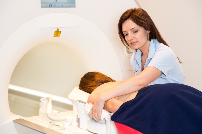
Emergency MRI of the Lumbar Spine – MRI is the gold standard diagnostic test for CES and must be ordered on an emergency basis — not as a routine or scheduled scan. Published clinical guidelines across multiple international spinal surgery associations are unanimous: urgent MRI is required for all patients presenting with new-onset urinary symptoms in the context of back pain or sciatica. The scan provides three-dimensional visualization of the spinal cord, nerve roots, and surrounding structures, confirming the presence and level of compression and identifying the underlying cause.
CT Scan or Myelogram When MRI Is Unavailable – If emergency MRI is not immediately available, a CT scan or CT myelogram can provide useful information about bony compression and spinal canal dimensions. These are secondary options — the absence of MRI availability does not justify delay in imaging. If necessary, the patient must be transferred to a facility with emergency MRI capability.
Surgical Referral Without Delay – Once imaging confirms cauda equina compression, the patient must be referred immediately to a spinal surgical unit. Emergency decompression surgery — typically laminectomy and discectomy — is the treatment for CES. Any avoidable delay between confirmed diagnosis and surgical intervention can cause permanent additional nerve damage.
Where the Diagnostic Process Goes Wrong
Despite the clarity of clinical guidelines on CES, diagnostic failures happen with troubling regularity. The most common failures that give rise to failure to diagnose malpractice claims include:
Common Questions About CES Diagnosis and Malpractice
Speak With a Florida CES Attorney Today
If your CES was misdiagnosed, your imaging was delayed, or your red flag symptoms were dismissed — and you went on to suffer permanent nerve damage that earlier intervention might have prevented — you may have a valid medical malpractice claim.
Lisa S. Levine, P.A. has handled and consulted on hundreds of Cauda Equina Syndrome cases across the United States. She understands the clinical standards that govern CES diagnosis and where those standards are breached.
Contact our office for a free, confidential case evaluation. We handle all CES cases on a contingency basis — you pay nothing unless we win.
Call us 24/7 at (954) 256-1820 or use the contact form on this page to get started.